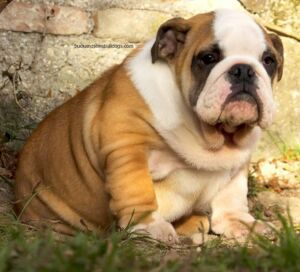
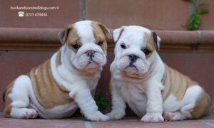
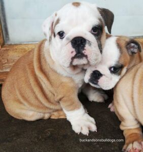
 The most frequent eye diseases in the English bulldog are: hypertrophy of the gland of the third eyelid, distichiasis, entropion, ectropion.
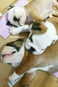
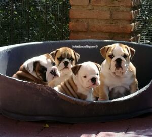
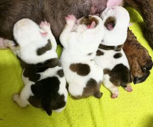
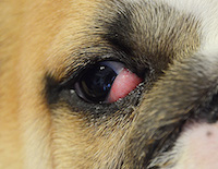
Hypertrophy of the gland of the third eyelid (Cherry Eye) is by far the most common eye disease in the bulldog. It occurs more frequently in young dogs, but sometimes (more rarely) it also occurs in adult dogs. The gland is normally present in all dogs but in some breeds, and among these the English bulldog, it tends to come out of its natural location due to a multiplicity of factors ranging from laxity of the ligament that holds it to the tendency to inflammation. The owner sees a sort of "cherry" in the inner corner of the eye. The gland usually comes out after an effort, wild games, or after the dog, exerting strength during a walk, is "pulled" for a long time with the leash. Sometimes, rarely, the gland returns to its place on its own during rest.
When the gland comes out it needs to be repositioned. Immediately, the owner can attempt a manual repositioning, exerting a slight downward pressure on the gland with the thumb to make it return to its seat (do not be afraid to cause pain to the dog, this maneuver is very simple and delicate; however, if this procedure scares you ask your vet for help). You can also try, initially, a drug treatment that usually gives poor results, but is still worth trying. In case of negative outcome of the treatment, it is necessary to proceed with the surgical repositioning. It is good to know that: in almost all cases this pathology is bilateral so, before proceeding with the surgery, it is always advisable to wait for the gland to come out in the other eye (this obviously to avoid having to operate the dog twice). In past years, the gland was mistakenly removed completely: the operation was performed without anesthesia and stitches. However, subsequent studies have shown that the gland is actually responsible for tearing of the eye and, if removed, its lack led to the onset (with the aging of the dog) of diseases such as keratoconjunctivitis sicca. The consequence, in these cases, is the administration (for the whole life of the dog) of eye drops that compensate for the lack of tearing, sometimes with poor results. Today, therefore, it is common practice to REPOSITION the gland, an operation that preserves its functionality.
The advice is always to contact an experienced ophthalmologist veterinarian, as the success of the operation is directly proportional to the surgeon's skill. Cases of relapse are very few if the veterinarian is experienced. The surgery is performed under total anesthesia and the post-operative course is quite simple. Unfortunately, in this type of pathology the selection has poor results because there is a strong breed predisposition which seems to be closely linked to the morphology of the English bulldog's head.